
Exosomes and Intercellular Tubular Connections
An entirely distinct form of unconventional secretion invokes the capture of cytoplasmic proteins and small RNA molecules into vesicles exported into the extracellular medium in cultured cells or the extracellular fluids in metazoan organisms. Extracellular vesicles (EVs) form in two distinct pathways. Microvesicles bud directly from the cell surface into the extracellular medium whereas a distinct subset of EVs are formed by membrane invagination into the interior of a late endosome to create a multivesicular body (MVB) which by fusion at the cell surface releases a bolus of EVs called exosomes. Much interest has developed around the evidence that EVs may serve as a means of intercellular communication in which proteins and small RNAs are delivered by fusion or uptake into target cells to mediate control of metabolism or gene expression. Of particular interest is the evidence that the microRNA content of EVs in the blood may change as a result of metastatic cancer and that EVs produced abundantly by tumor cells may communicate with other tissues in the body to create a premetastatic niche into which primary tumor cells may migrate to form a secondary metastatic event (11).
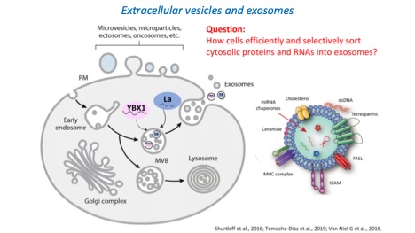
We have devised fractionation procedures to isolate distinct populations of EVs from normal and tumor cells grown in culture. Two vesicle types produced by a human breast cancer cell line are resolved on a buoyant density gradient and each contains a largely non-overlapping set of miRNAs (12). One vesicle type contains several highly selected mature miRNAs which we estimate are ~1000X enriched over their content in the cytoplasm of the tumor cell. A human embryonic cell line produces exosomes that also have several highly selected miRNAs but these differ from those enclosed within the selective pool of vesicles produced by the breast cancer cells (13, 14). These results suggest a cell type-specific high-fidelity sorting reaction to secrete only a subset of miRNAs from cells. This sorting may serve the purpose of selective disposal of certain miRNAs or the targeted delivery of selected miRNAs for control of gene expression in cells that take up and functionally incorporate the exosome content.
Little is known about the regulation of MVB fusion at the cell surface vs the lysosome. Recently we reported the surprising finding that MVB fusion at the cell surface is dramatically stimulated by conditions that damage the plasma membrane. Plasma membrane repair processes have been known for over a century and a repair process has been shown to involve the mobilization of mature lysosomes which fuse at point of rupture to patch damage at the cell surface (15). We reported that the same conditions and indeed some of the same Ca2+-binding proteins are involved in mobilizing MVBs and lysosomes for plasma membrane repair. These conditions greatly stimulate exosome secretion and may offer at least a partial explanation for the abundance of these vesicles in all bodily fluids.
Exosomes and EVs in general have been promoted as a vehicle for intercellular communication. Unfortunately, the evidence supporting this claim is indirect and very weak. For an EV/exosome to be an efficient cargo delivery vehicle, the luminal content of a vesicle must be discharged into the cytoplasm and possibly then into the nucleus of a target cell. This requires a membrane fusion reaction either at the cell surface or after the EV/exosome is internalized into an endosome. Enveloped RNA viruses which have membrane fusion protein catalysts integral to the virus particle serve as the precedent for this kind of delivery. No such membrane fusogen has been demonstrated in any of the published claims of functional cargo delivery by EV/exosomes. Indeed, the efficiency with which cargo is delivered by EVs has not been properly quantified and it remains possible to explain the many effects that have been reported by invoking a very inefficient delivery process.
We have developed two major findings with respect to efficient cargo delivery by EVs. We found that EVs secreted by neuroblastoma or mouse ES cells differentiating into neuronal precursor cells were taken up by and stimulated the rate of neuronal differentiation of mouse ES cells (16). EVs isolated from differentiating neurons were found to fairly selectively sort cyclinD1 and CDK4. Further, cyclinD1 was found to be rate-limiting and essential to speed the rate of differentiation of mouse ES cells. Using a proximity labeling approach, we showed that cyclinD1 in EVs reaches the nucleus in target cells to biotinylate two different nuclear proteins implicated in neuronal differentiation. Although this work did not define a fusogen or accurately quantify the efficiency of cyclinD1 delivery, the results clearly pointed to a mechanism for functional delivery of EV content to a target cell. This may be a specialized case where functional delivery of EV content serves an important role in stem cell differentiation.
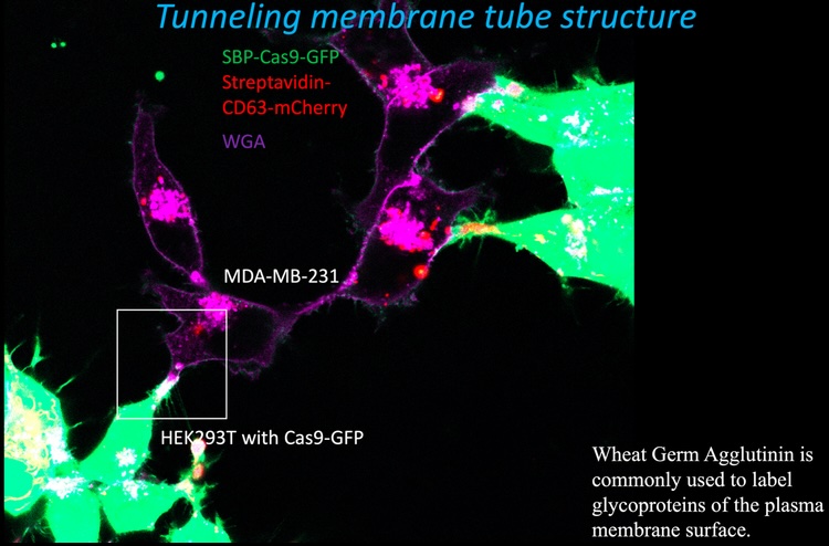
We devised a different assay to measure the efficiency of cargo delivery mediated by exosomes engineered to tether Cas9 indirectly to an endosome/exosome-specific membrane protein, CD63 (17). Exosomes isolated from HEK293 cells were 15-fold enriched in enzymatically active Cas9 and a gRNA expressed in the same cells. A recipient cell line was constructed using MDA-MB-231, a breast cancer cell line, expressing a crippled form of Nluc with a stop codon and a single nucleotide deletion introduced at the beginning of the reading frame just after a target sequence for the gRNA. Expression of Cas9 and the gRNA directly in the recipient cell resulted in error-prone repair at the site of ds cleavage sufficient to restore the reading frame and increase Nluc expression 60-100 fold. Exosomes isolated from donor cells were measurably internalized by recipient cells, but very little activation of Nluc gene expression was observed. In contrast, when donor and recipient cells were co-cultured with physical contact, substantial transfer of Cas9 and activation of Nluc expression in recipient cells was observed. Using fluorescence visualization and correlative light and electron microscopy (CLEM), we observed transfer mediated by open-ended membrane tubular connections between HEK2893 and MD-231 cells (fluorescence image below). The formation of such open-ended connections required the action of a membrane fusogen, syncytin, which is expressed in the recipient tumor cell line. Syncytin is normally expressed and functions only at the pre-implantation stage of embryonic development. However, its expression in tumors may suggest a role in tumor growth or metastasis, possibly through intercellular tubular connections or in exosomes secreted by tumor cells.